What is Poxvirus virus
Properties of Poxviruses
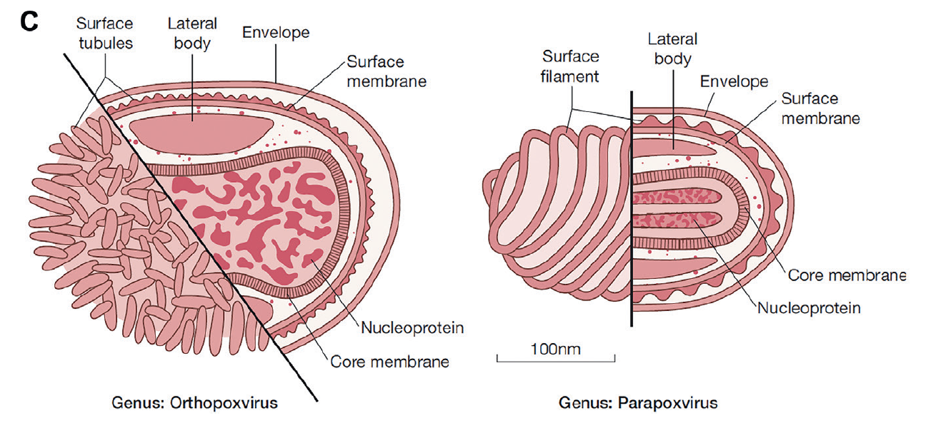
- Virions in most genera are brick-shaped virions, 250×200×200 nm, with an irregular arrangement of surface tubules. Virions of members of the genus Parapoxvirus are ovoid, 260×160 nm, with regular spiral arrangement of surface tubules
- Virions have a complex structure with a core, lateral bodies, outer membrane, and sometimes an envelope
- Genomes have the capacity to encode approximately 200 proteins, as many as 100 of which are contained within virions. Poxviruses, unlike other DNA viruses, encode all of the enzymes required for transcription and replication
- Cytoplasmic replication, enveloped virions released by exocytosis; non-enveloped virions released by cell lysis
Poxviruses are enveloped, large, double-stranded DNA viruses. Members of the poxvirus family infect a broad range of animal species, from insects to fish to birds to mammals. They pose a significant health threat for animals and humans. The best known is variola virus (VARV), the cause of smallpox, an extinct disease that claimed millions of victims and influenced human history.
Classification of Poxvirus
The family Poxviridae is divided into two subfamilies: Chordopoxvirinae (poxviruses of vertebrates) and Entomopoxvirinae (poxviruses of insects). The subfamily Chordopoxvirinae is divided further into nine genera, four of which (Orthipoxvirus, Parapoxvirus, Molluscipoxvirus, and Yatapoxvirus) contain viruses that cause human infections.
What’s the structure of Poxvirus virus
The poxvirus genome consists of a single molecule of linear double-stranded DNA varying in size from 130 kbp (parapoxviruses), to 280 kbp (fowlpox virus), and up to 375 kbp (entomopoxviruses). The genomes of vaccinia virus (191,636 bp) and many other poxviruses have been sequenced. Poxvirus genomes have covalent linkage joining the two DNA strands at both ends; the ends of each DNA strand have long inverted tandem repeated nucleotide sequences forming single-stranded loops (Figure).
Poxvirus genomes have the capacity to code for up to 200 proteins, as many as 100 of which are contained within virions. The genome can be divided functionally into a conserved central region coding for those proteins essential for virus replication (e.g., nucleic acid synthesis, virion structural component synthesis). Examples include a DNA-dependent DNA polymerase, DNA ligase, DNA-dependent RNA polymerase, enzymes involved in capping and polyadenylation of messenger RNAs and thymidine kinase. The two flanking regions toward the termini code for a large number of proteins that determine host range, virulence, and immunomodulation. In the case of vaccinia virus, these regions account for nearly half the genome.

Figure 1.Schematic diagram, genus Parapoxvirus. Part of the two diagrams shows the surface structure of an unenveloped virion, the other part shows a cross-section through the center of an enveloped virion.

Figure 2.Structure and organization of the genome of vaccinia virus.
How Poxvirus virus replicate
Most poxviruses, except for parapoxviruses and molluscum contagiosum virus, grow readily in cell culture. All produce pocks on the chorioallantoic membrane of embryonated hens’ eggs, the appearance of which was used to differentiate orthopoxviruses from each other before the introduction of molecular methods.
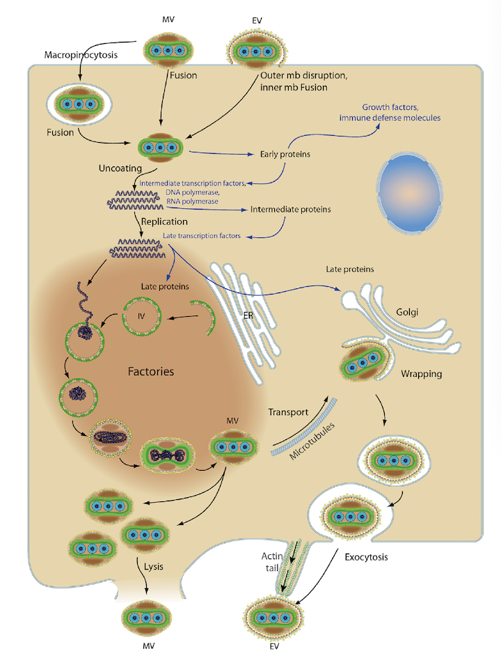
Unusually for a DNA virus, replication of poxviruses occurs entirely in the cytoplasm. To achieve this total independence from the host cell nucleus, poxviruses, unlike other DNA viruses, have evolved to code for all of the enzymes required for transcription and replication of the viral genome; several of these enzymes are carried in the virion itself. Virus entry into the target cell is by fusion of the virion with the plasma membrane or by endocytosis; after entry the viral core is released into the host cell cytoplasm

Fiture 3.The replication cycle of vaccinia virus.
Replication Process
1. Viral proteins attach to host cell membrane glycosaminoglycans (GAGs), and virions are endocytosed into the host cell. Alternatively, fusion with the plasma membranes can release cores into the host cytoplasm.
2. Early phase: Early genes are transcribed in the cytoplasm by viral RNA polymerase. Early expression begins at 30 minutes postinfection.
3. The core is completely uncoated as early expression ends; viral genome is now free in the cytoplasm.
4. Intermediate phase: Intermediate genes are expressed, triggering genomic DNA replication at approximately 100 minutes postinfection.
5. Late phase: Late genes are expressed from 140 minutes to 48 hours postinfection, producing all the structural proteins.
6. Assembly of progeny virions starts in cytoplasmic viral factories, producing a spherical immature particle (IV). This virus particle matures into the brick-shaped intracellular mature virion.
7. Intracellular MVs can be released after cell lysis, or can acquire a second double membrane from the trans-Golgi and bud as external enveloped virions (EV).
Reference:
- Fenner and White's Medical Virology 5th Edition
Echo Biosystems is committed to delivering high-quality proteins to support your scientific research. We have developed a series of high-quality poxvirus proteins. Please feel free to contact us if youo have any questions or customized requirement
- Product Name
- Organism
- Tag (Tag info)
- Expression Host (Source)
-
Recombinant Vaccinia virus Protein L1(VACWR088),partialVaccinia virus (strain Western Reserve) (VACV) (Vaccinia virus (strain WR))N-terminal 6xHis-taggedE.coli
Recombinant Vaccinia virus Protein L1(VACWR088),partial
-
Recombinant Vaccinia virus Protein L1(VACWR088),partialVaccinia virus (strain Western Reserve) (VACV) (Vaccinia virus (strain WR))N-terminal 10xHis-tagged and C-terminal Myc-taggedBaculovirus
Recombinant Vaccinia virus Protein L1(VACWR088),partial
-
Recombinant Vaccinia virus Protein B5(PS/HR),partialVaccinia virus (strain Western Reserve) (VACV) (Vaccinia virus (strain WR))N-terminal 10xHis-tagged and C-terminal Myc-taggedBaculovirus
Recombinant Vaccinia virus Protein B5(PS/HR),partial
-
Recombinant Vaccinia virus Protein B5(PS/HR),partialVaccinia virus (strain Copenhagen) (VACV)N-terminal 6xHis-taggedYeast
Recombinant Vaccinia virus Protein B5(PS/HR),partial
-
Recombinant Vaccinia virus Protein K3 (VACWR034)Vaccinia virus (strain Western Reserve) (VACV) (Vaccinia virus (strain WR))N-terminal 6xHis-taggedYeast
Recombinant Vaccinia virus Protein K3 (VACWR034)
-
Recombinant Vaccinia virus 14 kDa fusion protein(A27L)Vaccinia virus (strain Copenhagen) (VACV)N-terminal 6xHis-sumostar-taggedYeast
Recombinant Vaccinia virus 14 kDa fusion protein(A27L)
-
Recombinant Vaccinia virus Truncated plaque-size/host range protein(PS/HR)Vaccinia virus (strain LC16m8) (VACV)N-terminal 6xHis-taggedYeast
Recombinant Vaccinia virus Truncated plaque-size/host range protein(PS/HR)
-
Recombinant Variola virus 14 kDa fusion protein(A27L)Variola virus (isolate Human/India/Ind3/1967) (VARV) (Smallpox virus)N-terminal 6xHis-taggedYeast
Recombinant Variola virus 14 kDa fusion protein(A27L)
-
Recombinant Variola virus 14 kDa fusion protein(A27L)Variola virus (isolate Human/India/Ind3/1967) (VARV) (Smallpox virus)N-terminal 10xHis-taggedYeast
Recombinant Variola virus 14 kDa fusion protein(A27L)
-
Recombinant Vaccinia virus Protein L1(VACWR088),partialVaccinia virus (strain Western Reserve) (VACV) (Vaccinia virus (strain WR))N-terminal 6xHis-taggedYeast
Recombinant Vaccinia virus Protein L1(VACWR088),partial
